Differential interference contrast microscopy (DIC)
DIC works by separating a polarised light source into two beams which take slightly different paths through the sample. Where the length of each optical path (i.e. the product of refractive index and geometric path length) differs, the beams interfere when they are recombined. This gives the appearance of a three-dimensional physical relief corresponding to the variation of optical density of the sample, emphasising lines and edges though not providing a topographically accurate image.

1. Unpolarised light enters the microscope and is polarised at 45°.
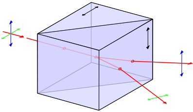 2. The polarised light enters the first Nomarski-modified Wollaston prism and is separated into two rays polarised at 90° to each other, the sampling and reference rays.
3. The two rays are focused by the condenser for passage through the sample. These two rays are focused so they will pass through two adjacent points in the sample, around 0.2 μm apart.
4. The rays travel through the different, adjacent, areas of the sample. They will experience different optical path lengths where the areas differ in refractive index or thickness. This causes a change in phase of one ray relative to the other due to the delay experienced by the wave in the more optically dense material.
5. The rays travel through the objective lens and are focused for the second Nomarski-modified Wollaston prism.
6. The second prism recombines the two rays into one polarised at 135°. The combination of the rays leads to interference, brightening or darkening the image at that point according to the optical path difference.
6. The second prism recombines the two rays into one polarised at 135°. The combination of the rays leads to interference, brightening or darkening the image at that point according to the optical path difference.
Image is produced by brightfield of 2 adjacent images + interferance by path differences. 3D effect is artificial. Detects different optical density. Used for live cell visualisation, don't need stain. No halos.

No comments:
Post a Comment