Fluorescence microscopy
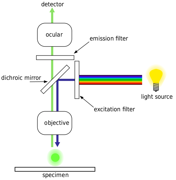 Epifluorescence microscopy is a method of fluorescence microscopy that is widely used in life sciences. The excitatory light is passed from above (or, for inverted microscopes, from below), through the objective and then onto the specimen instead of passing it first through the specimen. (In the latter case the transmitted excitatory light reaches the objective together with light emitted from the specimen). The fluorescence in the specimen gives rise to emitted light which is focused to the detector by the same objective that is used for the excitation. A filter between the objective and the detector filters out the excitation light from fluorescent light. Since most of the excitatory light is transmitted through the specimen, only reflected excitatory light reaches the objective together with the emitted light and this method therefore gives an improved signal to noise ratio.
Confocal microscopy

A confocal microscope uses point illumination and a pinhole in an optically conjugate plane in front of the detector to eliminate out-of-focus information. As only light produced by fluorescence very close to the focal plane can be detected the image resolution, particularly in the sample depth direction, is much better than that of wide-field microscopes. However as much of the light from sample fluorescence is blocked at the pinhole this increased resolution is at at the cost of decreased signal intensity so long exposures are often required.
Good for constructing 3D models. Good for looking through thick slices.

No comments:
Post a Comment